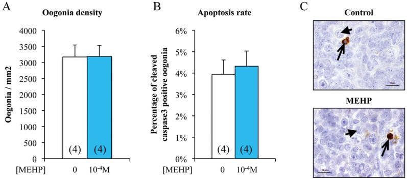
Figure 3. In vitro exposure to MEHP does not affect the number or apoptosis rate of germ cells in human fetal ovaries.
Ovaries from 7 to 12 week/old human embryos were cultured with or without 10−4 M MEHP for three days. At the end of the culture period, they were fixed with Bouin’s solution and stained with hematoxylin and eosin. Oogonia density (number of germ cells per mm2 tissue) was measured based on the morphological analysis (A) and germ cell apoptosis rate (B) based on the expression of cleaved Caspase-3 (brown) (C). Histograms represent the mean ± SEM of four different ovaries from different fetuses (as indicated in the columns). Arrowheads indicate oogonia and arrows cleaved Caspase- 3 positive oogonia. Bar, 15 µm.

