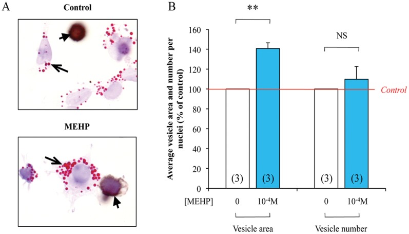
Figure 6. Quantification of lipid vesicle in dispersed testis cells following 72.
h of 10−4 M MEHP exposure. Human fetal testes were treated, or not, with 10−4 M MEHP for 3 days in organotypic cultures. Testes were then dissociated and Oil Red O (Red, arrows) and M2A (Brown, arrowheads) staining were performed (A). In the somatic cells, quantification of lipid vesicle number per nuclei and average vesicle area was performed using Image J software (B). Control are set to 100% and treated are expressed in percentage of the control. The histograms are the mean ± SEM of 3 independent experiments from different fetuses (as indicated in the respective column). NS = Not Significant, **p<0.01 in paired t-tests as recommended when comparing few samples.

