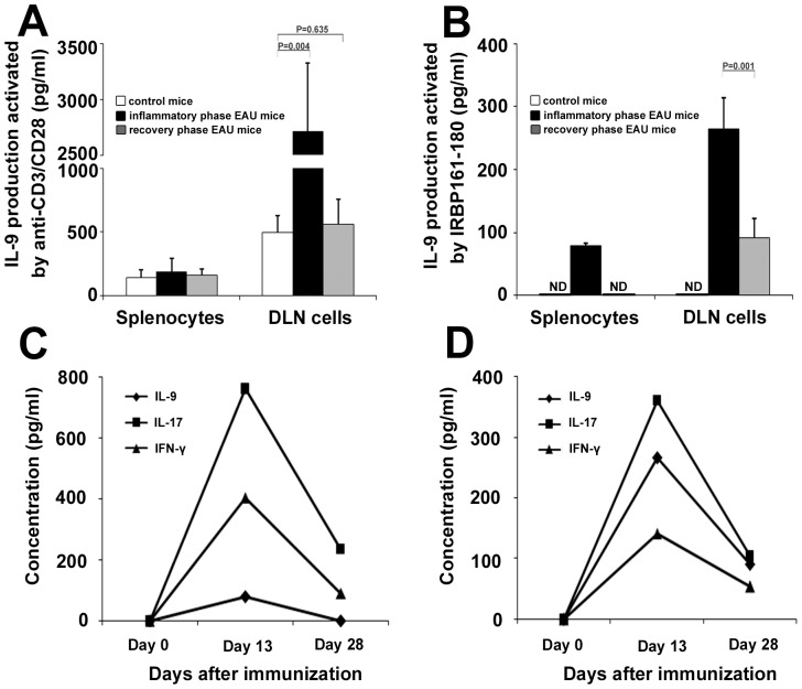
Figure 1. The expression of IL-9 in the EAU mice and the control mice.
Splenocytes and DLN cells, obtained from the EAU mice (inflammatory phase and recovery phase) or control mice (n = 5 per group), were activated with anti-CD3/D28 (1 µg/ml) (A) or IRBP161–180 (20 µg/ml) (B) for 3 days. IL-9 was analyzed by ELISA. Splenocytes (C) and DLN cells (D), obtained from the immunized mice on indicated time points, were stimulated with IRBP161–180 (20 µg/ml) for 3 days, and the supernatants were collected for measuring IL-9, IL-17 and IFN-γ. Data are representative of three independent experiments. ND: not detected.

