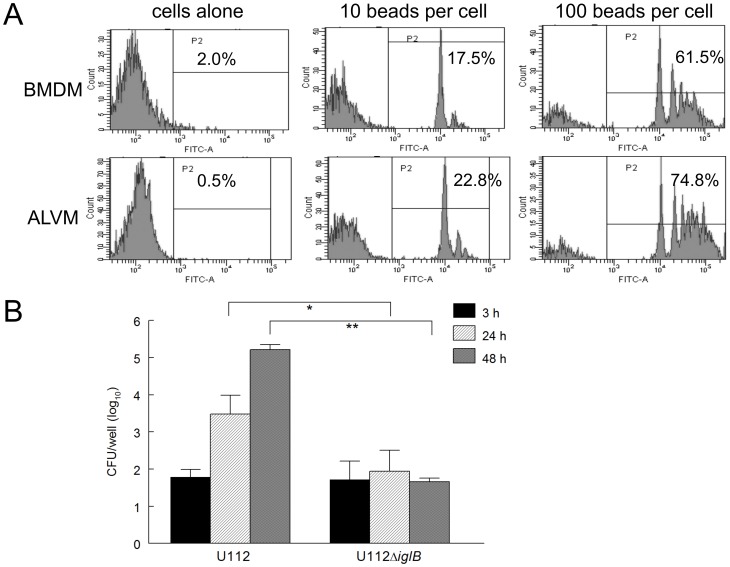
Figure 2. The phagocytic capacity of F344 alveolar macrophages.
A) Bone marrow derived macrophages (BMDM) or alveolar macrophages (ALVM) were seeded (1×10⁶ cells/well) into 6-well plates and allowed to adhere. Fluorescent beads were added to each cell type at either 10 or 100 beads/cell and incubated for 2 hr to allow for phagocytosis. Cells were washed to remove unphagocytosed beads and then stained for flow cytometry analysis. B) ALVM (2×10⁵ cells/well) were seeded, allowed to adhere, and infected for 2 hr with 10 MOI of either U112 or the live attenuated defined mutant strain U112ΔiglB. Cells were subsequently treated with gentamicin for one hour to kill any remaining extracellular bacteria and incubated at 37°C for 48 hr. At defined time points (3, 24, and 48 hr), cells were lysed with 0.2% (w/v) deoxycholate and dilution plated to enumerate intracellular bacteria. Differences between U112 and U112ΔiglB at 24 and 48 hr were significant (*p<0.05 and **p<0.005, respectively). Results are representative of two separate experiments.

