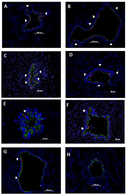
Figure 3.

Depletion of epithelial CCSP occurs in BOS but is preserved in BOS-free and normal donor tissues: Tissue is co-immuno-stained for CCSP (red) and AT (green) and nuclear counter stained with DAPI (blue). White arrow heads show areas of CCSP expression throughout an airway. CCSP and AT were assessed in the airways of BOS-free donor tissue (A, B), BOS-free transplant tissue (C, D), early BOS tissue (E, F), and advanced BOS tissue (G, H). Representative airways showing normal CCSP expression in BOS-free donor tissue (A, B) and BOS-free transplant tissue (C, D). Representative airways demonstrating the depletion of CCSP in BOS 1(E) and BOS 2 (F). Similar CCSP loss in the airways of advanced BOS 3 (G,H). CCSP expression in the BOS free donor tissue is very similar to BOS free transplant tissue, while early BOS tissue and advanced BOS tissue show significant CCSP loss. Images A–D, F are presented at 200x, image E is presented at 400X.
