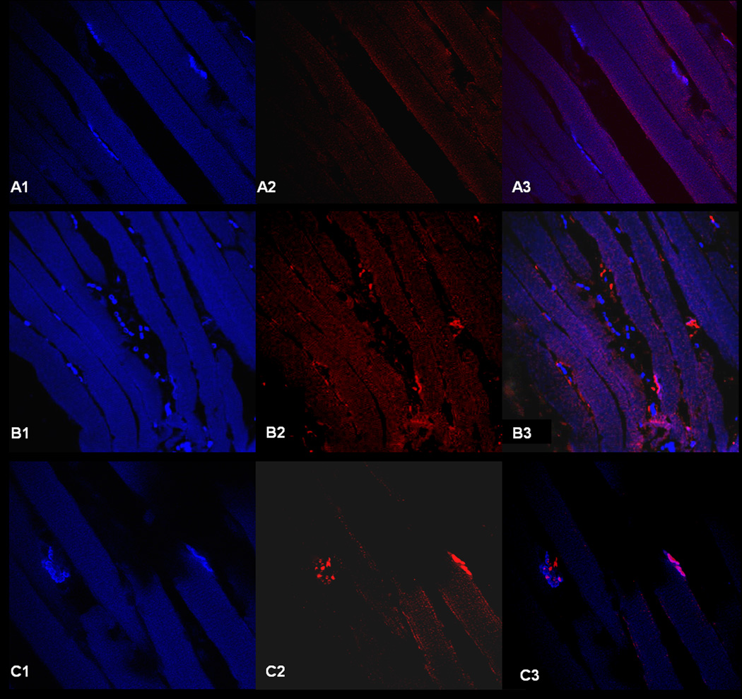
Figure 9. Motor Endplate to Nerve Contact.
Representative high power (60X) confocal microphotographs of immunohistochemistry slides with bungarotoxin (blue) demarcating the motor endplates along the myofibers (left), and the middle figures demonstrating staining with the neuronal specific marker, beta III tubulin (red). On the right, the two images are merged; those sites with colocalization of the bungarotoxin (blue) and beta III tubulin (red) represent motor endplates with nerve contact (reinnervation), with the areas of bungarotoxin alone (blue) representing denervated motor endplates. Figures A1–A3 demonstrates a section series classified as denervated (<33% of motor endplates with nerve contact), while B1-3 demonstrates a partially reinnervated section (33–66% of motor endplates with nerve contact), and C1-C3 demonstrates a strongly reinnervated section (>66% of motor endplates with nerve contact). Note the high variability in the number of motor endplates between sections, with series B1-B3 having the greatest number of motor endplates within the section.

