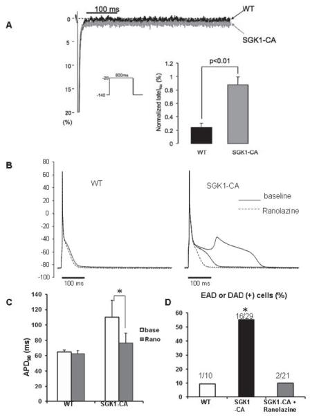
Figure 5.

SGK1 activation increases INaL while ranolazine normalizes APD and suppresses afterdepolarizations in SGK1-CA cardiomyocytes. (A) Representative normalized INaL (% of peak) superimposed for WT and SGK1-CA cardiomyocytes. Inset shows that normalized INaL was larger in SGK1-CA (n=6) than in WT cardiomyocytes (n=5). (B) Superimposed APs in cardiomyocytes from WT and SGK1-CA mice before (baseline: solid line) and after ranolazine (dotted line) at 0.5Hz pacing. Ranolazine (1μmol/L) normalized APD and suppressed EAD in SGK but did not affect APs in WT myocytes. (C) Ranolazine (1μmol/L) normalized APD90 in SGK1-CA cardiomyocytes (*p<0.05, repeated measures ANOVA), but did not affect APD90 in WT cardiomyocytes. (D) EADs and DADs were more frequent in SGK1-CA compared with WT cardiomyocytes. Ranolazine (1 μmol/L) reduced after-depolarizations in SGK1-CA cardiomyocytes (*p<0.05 by χ2 test) to levels seen in WT cardiomyocytes.
