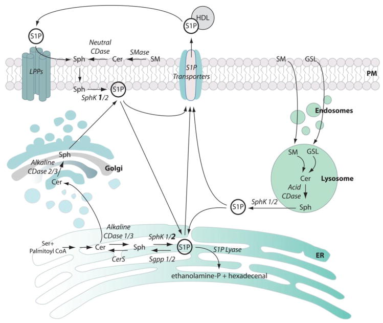
FIGURE 1. Cellular S1P metabolism.
Cellular synthesis and degradation of S1P involves multiple enzymes, some expressed as various isoforms with different biochemical properties and cellular locations, indicating a highly complex metabolism. S1P synthesis by Sphk1 and 2 can occur after the degradation of ceramide, which takes place in the ER and in the Golgi after de novo synthesis, or in the lysosomes and at the plasma membrane, during catabolism of sphingomyelin and glycosphingolipids. Sphk1 activity (bold) predominates at the plasma membrane, while Sphk2 activity (bold) predominates for ER associated S1P metabolism. Within the cell, S1P may move rapidly between cell compartments. Intracellular S1P can be degraded by S1P lyase, dephosphorylated by S1P phosphatases to recycle the sphingoid base for ceramide synthesis, or else secreted. S1P is transported outside the cell by members of the ABC family of transporters and by Spns2. Once in the extracellular space, S1P can be dephosphorylated by a group of lipid phosphatases, LPPs, liberating sphingosine, which can be rephosphorylated back to S1P. Sph, sphingosine; Cer, ceramide; ethanolamine-P, ethanolamine phosphate; SM, sphingomyelin; GSL, glycosphingolipids; Ser, serine; LPPs, lipid phosphatases; CerS, ceramide synthase; CDase, ceramidase; SMase, sphingomyelinase; PM, plasma membrane.

