Figure 4.
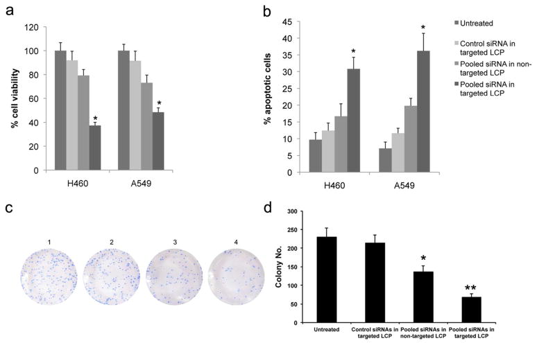
In vitro oncogenes silencing inhibits NSCLC proliferation and induces apoptosis. (a) Cell viability was measured by MTT assay after treatment with different LCP for 48 h. (b) Histogram showing the quantification of apoptotic cells treated with different LCP for 48 h. (c) Representative colony formation assay of NSCLC cells treated with different formulation. 1) untreated; 2) control siRNA in targeted LCP; 3) pooled siRNA in non-targeted LCP; 4) pooled siRNA in targeted LCP. All colonies were stained with crystal violet. (d) Histogram showing the quantification of colony formation efficiency. Columns, mean (n = 3); bars, SD. *, p < 0.05; **p < 0.01.

