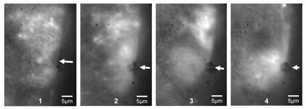
FIG. 8.
Fluorescent image sequence showing microstreaming around a dark circular microbubble pointed out by the arrows in each frame. Frame 1 shows the field of non-echogenic liposomes before ultrasound application. Frame 2 shows the microstreaming flow field starting at the beginning of ensonation. The diameter of the flow field is 6 times larger than the diameter of the microbubble itself and can pull in larger clusters of fluorescent liposomes as seen in frames 3 and 4.

