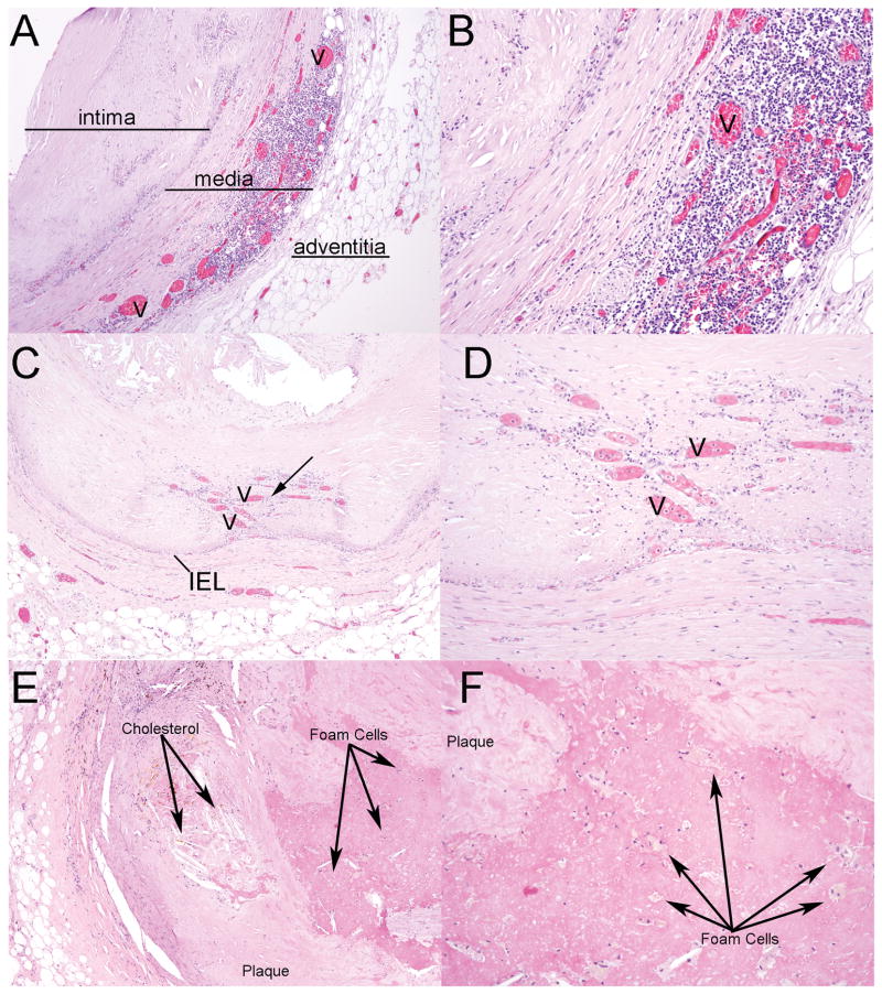
Fig. 3.
Histological changes in the medial and intimal layers of coronary vessels with atherosclerosis. (A) Image of neovascularization (v) and inflammatory cells within the muscular layer of the coronary vessel (hematoxylin and eosin, original magnification × 40). (B) Higher-power image (original magnification × 100). (C) Image of neovascularization (arrow) that is in the intimal region and is adjacent to dystrophic calcification (original magnification × 40). (D) Higher-power image (original magnification × 100). (E) Photomicrograph of cholesterol accumulation within an atherosclerotic lesion with calcification (original magnification × 40). (F) Higher power image highlighting the presence of foam cells within the plaque (original magnification × 100). Cholesterol and foam cells indicated as labeled. IEL=internal elastic lamina.

