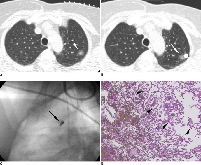
Fig. 1.
Percutaneous localization of part-solid nodule in left upper lobe using barium sulfate in 68-year-old woman.
A. Appropriate barium injection site and needle route were determined using pre-procedural CT. Note part-solid nodule (arrowhead) in left upper lobe and radiopaque grid made of metallic wires on chest wall. B. Barium sulfate suspension was injected adjacent to target nodule (arrow) and appears as radiopaque ball on post-procedural CT image. C. Intraoperative fluoroscopic image shows radiopaque barium ball (arrow) in left upper lung zone. D. Injected barium sulfate appears as brownish materials dispersed within alveolar space. Note nodule seen as part-solid nodule on CT (arrowheads) near injected barium materials. There are acute inflammatory reactions containing many neutrophils, histiocytes and some eosinophils around injected barium material (original magnification, × 200; Hematoxylin-Eosin stain). Target nodule was confirmed as adenocarcinoma-in-situ, pathologically.

