Fig. 2.
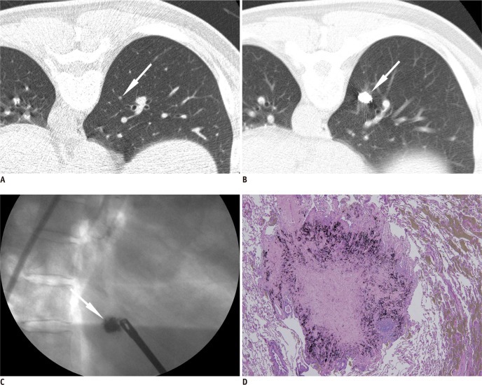
Percutaneous transthoracic localization of tiny solid nodule prior to VATS using barium sulfate in 56-year-old man with medical history of colon cancer.
A. Tiny solid nodule was found on routine follow-up CT scan (arrow). B. Post-procedural CT image shows injected barium ball encroaching target nodule (arrow). C. Intraoperative fluoroscopic image shows radiopaque barium ball (arrow) in right lower lung zone. D. Brownish barium sulfate is shown around target nodule confirmed as intrapulmonary lymph node, pathologically. VATS = video-assisted thoracoscopic surgery

