Fig. 3.
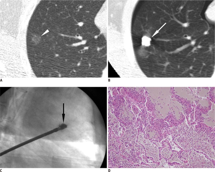
Preoperative localization of pure ground-glass nodule using barium sulfate in right upper lobe found in 62-year-old man.
A. Thin-section chest CT scan shows 1 cm pure ground-glass nodule (arrowhead) in right upper lobe. B. Injected barium near target nodule is clearly demonstrated on post-procedural CT (arrow). Note small amount of pneumothorax which occurred during localization procedure. C. Intraoperative fluoroscopic imaging shows radiopaque barium ball (arrow) in right upper lung zone. D. Scattered brownish colored barium with acute inflammation reaction is shown. Lesion was confirmed as atypical adenomatous hyperplasia, pathologically.

