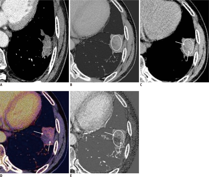
Fig. 2.
Hemorrhagic tumor response detected on dual-energy CT in 66-year-old man with lung adenocarcinoma.
A. Previous baseline enhanced CT image shows 35 mm-sized primary tumor with net enhancement of 48 HU (subtraction of CT attenuation value on nonenhanced image from that of enhanced image; 68 minus 20 HU) in left lower lobe. B. Enhanced weighted-average image obtained from dual-energy CT after chemotherapy shows about 28% increase in size of primary tumor and also increased CT attenuation value (up to 82 HU, circular regional of interest [ROI]). Thus, tumor response was assessed as progressive disease on basis of traditional RECIST 1.1. C. In contrast, virtual nonenhanced image shows primary cancer CT attenuation value of 53 HU (circular ROI), suggesting hemorrhagic component (arrows). D, E. Color coded (D) and grayscale (E) iodine-enhanced images show tumor (arrows) attenuation value of 29 HU, declined value by 40% of net enhancement calculated from previous baseline chest CT. Therefore, tumor response was assessed as partial response by Choi's criteria based on dual-energy CT.

