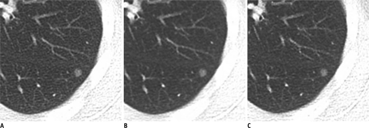
Fig. 3.
Low dose chest CT without enhancement in 37-year-old woman (BMI: 23 kg/m2) with small ground glass nodules.
A-C. Transverse CT images with (A) full radiation dose FBP in B70f kernel and 5 mm slice thickness, (B) full radiation dose IRIS in I70f kernel and 5 mm slice thickness, and (C) half radiation dose IRIS in I70f kernel and 5 mm slice thickness. Objective noise measured in saline bag was 67.8 HU, 41.5 HU and 58.2 HU, respectively (not shown). BMI = body mass index, HU = Hounsfield unit, FBP = filtered back projection, IRIS = iterative reconstruction in image space

