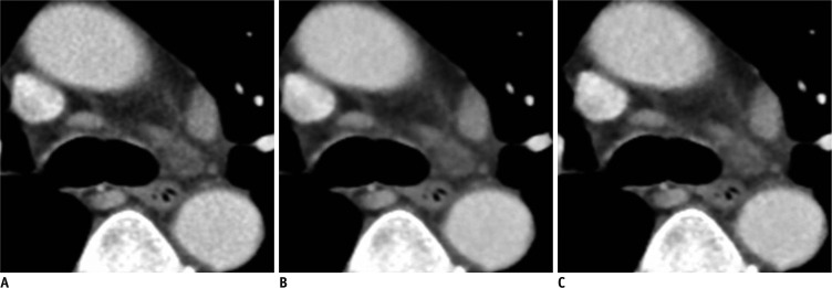
Fig. 4.
Standard dose chest CT with enhancement in 75-year-old man (BMI: 21 kg/m2) with small lymph nodes in mediastinum.
A-C. Transverse CT images with (A) full radiation dose FBP in B30f kernel and 5 mm slice thickness, (B) full radiation dose IRIS in I30f kernel and 5 mm slice thickness, and (C) half radiation dose IRIS in I30f kernel and 5 mm slice thickness. Objective noise measured in saline bag was 59.7 HU, 34.5 HU and 48.5 HU, respectively (not shown). Margins of mediastinal structures were slightly more blurred in (B) and (C) than in (A) due to excessive smoothing effect of IRIS image. BMI = body mass index, HU = Hounsfield unit, FBP = filtered back projection, IRIS = iterative reconstruction in image space

