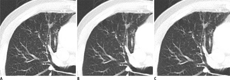
Fig. 1.
Standard dose contrast enhanced-chest CT in 77-year-old man (BMI 22 kg/m2) with sequalae of previous inflammation in right upper lobe.
A-C. Transverse CT images at 5 mm thickness with full radiation dose reconstructed with (A) filtered back projection (FBP) and (B) image reconstruction in image space (IRIS), and (C) with half radiation dose reconstructed with IRIS. When two readers compared F-FBP (A) and F-IRIS (B) images as reconstruction effect, their preference scores were 5 and 4 for lung 5 mm images, 2 and 3 for mediastinum, 5 and 3 for lung 1 mm images, and 5 and 4 for overall images (not shown). When two readers compared F-FBP (A) and H-IRIS (C) images as reconstruction effect and radiation dose effect, readers' preference scores were 4 and 3 for lung 5 mm images, 3 and 3 for mediastinum, 4 and 4 for lung 1 mm images, and 4 and 3 for overall images (not shown). BMI = body mass index, F-FBP = full dose image with filtered back projection, F-IRIS = full dose image with iterative reconstruction in image space, H-IRIS = half dose image with iterative reconstruction in image space

