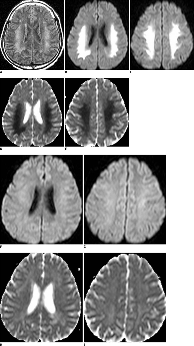
Fig. 1.
Brain MRI in 36-year-old man with right facial palsy and motor weakness.
Brain MRI performed upon admission. Initial FLAIR reveals confluent high signal intensities of cerebral white mater, sparing cortex and basal ganglia (A). These lesions show high signal intensities on DWI and decreased ADC values (B-E). FLAIR = fluid-attenuated inversion recovery, DWI = diffusion-weighted imaging, ADC = apparent diffusion coefficient. Follow-up magnetic resonance (MR) scan obtained after 7 days, when symptoms had improved. MR images show that lesions, which showed abnormal signal intensities, are completely resolved (F-I).

