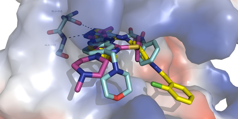
Fig. 1.
The crystallographic binding modes of three AKIs (in sticks, cyan—AT-9283, PDB (protein data bank) code 2W1E; yellow—bisanilinopyrimidine-based AKI, PDB code 3H0Y, violet—VX-680, PDB code 3E5A in the Aurora kinase A binding cleft (shown as surface). Specific hydrogen bonds to the backbone of residues Glu-211 and Ala-213 in the hinge region are shown by dotted lines. Color coding: oxygen—red, nitrogen—blue, chlorine—green, carbon—different colors. The figure was prepared using PyMol, ver. 0.99 [31]

