Figure 1.
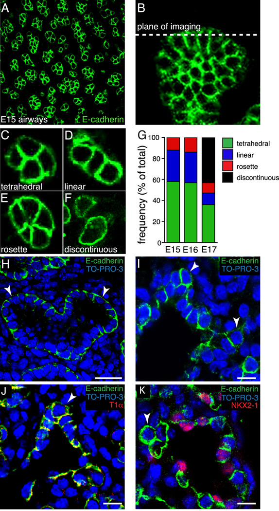
Epithelial cell orientation at the tips of E15, E16, and E17 fetal mouse lung airways. (A). Representative confocal image of E-cadherin expression in E15 mouse whole lung lobe. (B). Z-projection of E-cadherin expression in whole lung lobe showing the plane of imaging used for measuring cell orientation. (C–F) E-cadherin immunolabeling and confocal imaging revealed four distinct epithelial cell orientations; tetrahedral (C), linear (D), rosette (E), and discontinuous (F). (G) Similar frequencies of epithelial orientations in E15 and E16 airways (n = 230, 201, respectively). At E17 (n = 347), over 40% of the airways contained epithelia in a discontinuous orientation, with remaining airways distributed among the three orientations observed at E15 and E16. (H, I). E-cadherin immunolabeling of E15 mouse lung frozen sections demonstrated distinct epithelial monolayer surrounding the lumen of branched airways (H). E17 epithelial cells at the tips of branching airways (arrowheads) did not maintain a monolayer, but appeared to invade the mesenchyme (I). E18 tip epithelial cells (arrowheads) expressed type I epithelial cell marker, T1α (J), but lacked expression of NKX2-1, type II epithelial cell marker (K). (H) Scale bar, 25 µm. (I–K) Scale bar, 10 µm.

