Figure 3.
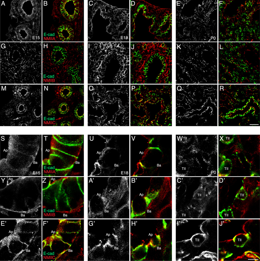
Non-muscle myosin II (NM II) isoform expression in the developing fetal mouse lung. Confocal images of E15 (B, H, N, T, Z, F’), E18 (D, J, P, V, B’, H’), and P0 (F, L, R, X, D’, J’) mouse lung frozen sections immunostained for E-cadherin (green) and NM II-A, NM II-B, or NM II-C (red). Single channel confocal images of NM II isoform immunostained mouse lung frozen sections at E15 (A, G, M, S, Y, E’), E18 (C, I, O, U, A’, G’), and P0 (E, K, Q, W, C’, I’). (A–F, S–X) NM II-A was present in both the fetal lung mesenchyme and epithelium at E15 and E18, but localized away from type II alveolar epithelial cells at P0. (G–L, Y–D’) NM II-B localized to the fetal lung mesenchyme. (M–R, E’–J’) NM II-C co-localized with E-cadherin along the apical and basolateral surfaces of fetal airway epithelial cells and was expressed in both alveolar type I and type II epithelial cells (R, J’). (A–R) Scale bar, 25 µm. (S–J’) Scale bar, 5 µm.

