Figure 7.
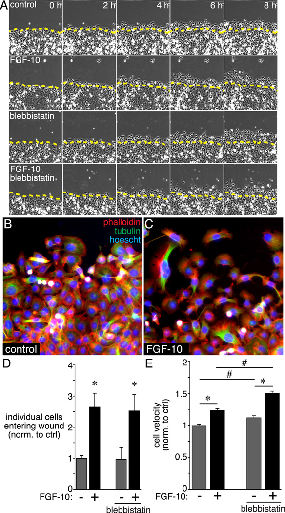
Effects of blebbistatin on FGF-10-stimulated epithelial cell migration. MLE-12 lung epithelial cells were grown to confluence and then scratched to produce an artificial wound. Following wounding, cells were treated with FGF-10 +/− blebbistatin and imaged by time-lapse microscopy for 8 h. (A) Individual still images obtained by phase contrast microscopy show migration of cells into the wound. Images were obtained every 20 min, and representative images from 0 h, 2 h, 4 h, 6 h, and 8 h are shown. (B, C) Control and FGF-10 treated MLE-12 cells were labeled with FITC-conjugated anti-tubulin (green) and Alexa594 phalloidin (red) at 8 h following wounding. (D) FGF-10 treatment increased the number of cells migrating into the wound and losing contact with the existing monolayer (* P < 0.05, n = 9). Blebbistatin treatment did not have any effect on the number of cells entering the wound. (E) FGF-10 increased MLE-12 cell velocity in wounded cultures (* P < 0.05, n = 120). Blebbistatin treatment increased velocity in both control and FGF-10-treated cells (# P < 0.05, n = 120).

