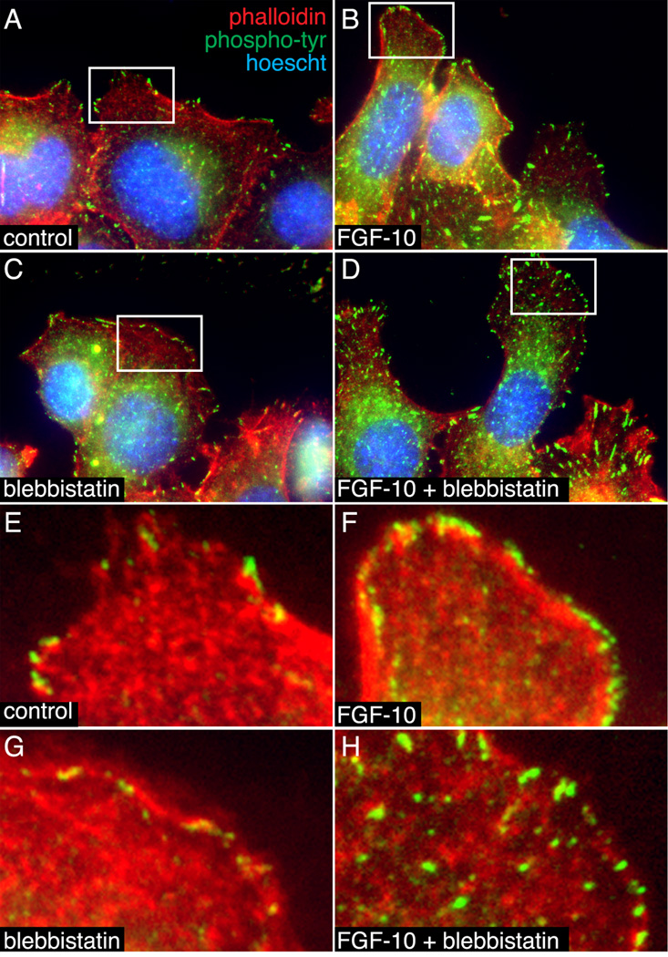
Figure 8.
Blebbistatin altered the subcellular localization of phosphotyrosine staining in MLE-12 cells. (A–D). MLE-12 cells treated with FGF-10 +/− blebbistatin were labeled with FITC-conjugated anti-phophotyrosine (green) and Alexa594-phalloidin (red). Nuclei were labeled with Hoescht dye (blue). (B). FGF-10 increased phosphotyrosine staining at the cell periphery. (C, D). Blebbistatin decreased the size of phosphotyrosine-positive focal contacts in control cells and caused a more generalized distribution following FGF-10 treatment. Lower magnification images were taken by widefield fluorescence microscopy (A–D). Higher magnification images obtained by laser scanning confocal microscopy are included in panels (E–H).

