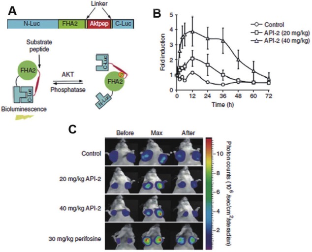
Figure 4.
A: Schematic representation of mechanism of action of Bioluminescent Akt reporter (BAR): BAR consist of an Akt consensus substrate peptide (Aktpep) and phospho-amino acid binding domain (FHA2) which are flanked by the N-terminal (N-Luc) and C-terminal (C-Luc) of the firefly luciferase reporter molecule respectively (upper panel). Akt dependent phosphorylation of the Aktpep domain induces interaction with the FHA2 domain (right) resulting minimal bioluminescence activity. In absence of Akt activity, the N-Luc and C-Luc domains reassociate restoring bioluminescence activity (left) (lower panel) B: Graphical representation of imaging Akt inhibition: D54-BAR (WT) stably transfected cells were implanted subcutaneously into nude mice. Tumor-specific bioluminescence activity was monitored 4 weeks after implantation, when tumors reached about 40–60 mm3 in size. Bioluminescence activity before treatment (time 0) and in response to treatment with vehicle control (20% DMSO in PBS) and API-2, Akt inhibitor (20 mg/kg or 40 mg/kg) was monitored at various times Bioluminescence activity remained flat over a 48-h period in vehicle-treated mice. In contrast, in mice treated with API-2 (40 mg/kg), bioluminescence activity increased within first 2h and reached a peak within 6h after treatment as compared to the small increase in activity on treatment with API-2 (20 mg/kg) The result suggest that dose in excess of 40 mg/kg of API-2 are required for maximal inhibition. C: Representative bioluminescence images of mice: Tumor bearing mice were treated with vehicle control (20%DMSO), API-2 (20mg/kg and 40mg/kg) and perifosine (PI3K inhibitor) (30 mg/kg). Images of representative mice are shown before treatment, during maximal luciferase signal upon treatment (Max), and after treatment. (Adapted from 76 with permission).

