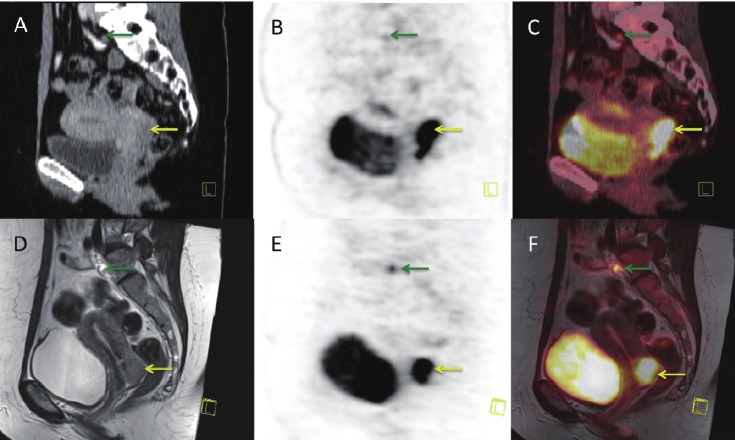
Figure 2.
Sagittal PET/CT and PET/MR images of a patient with cervical cancer (yellow arrow) and one pathological pelvic lymph node (green arrow): A: CT-scan, B: FDG-PET scan performed on the PET/CT scanner, C: Fused PET/CT image –note the mismatch between the bladder on CT and PET due to the difference in uptake time, D: MR-scan, T2 weighted, E: PET scan acquired simultaneous with MR, F: fused PET/MR image –note the perfect fit with bladder on MR and PET.

