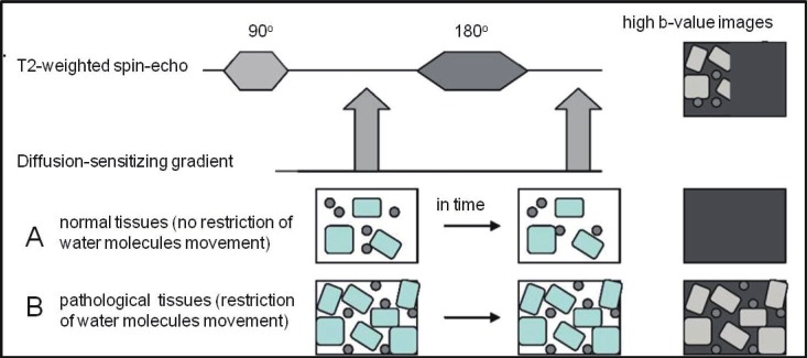
Figure 3.
Illustration of the principle behind diffusion-weighted MRI: During diffusion-weighted MRI two symmetric diffusion-sensitizing gradients are applied (1, 2) around the 180° refocusing pulse in the standard T2-weighted spinecho sequence. After the first (1) diffusion gradient, resonance frequencies of effected tissues (in particular, water molecules) will be changed, so that dephasing of the transverse magnetization will happen. Re-applying the same gradient for the same duration but of opposite polarity (2), “rephasing” of the transverse magnetization will emerge for stationary water molecules and there will be no significant change in the measured signal intensity (B). This second (2) diffusion gradient will not influence moving water molecules (A) because they have altered their spatial positions, so that incomplete rephrasing of the transverse magnetization will happen, which is displayed as a signal loss.

