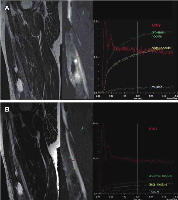
Figure 6.
Chemotherapy monitoring with dynamic contrast-enhanced (DCE) MRI: A patient with recurrent myxoid liposarcoma of the soft tissues in the right thigh before (A) and after (B) chemotherapy. A: Sagittal T2-weighted TSE image before treatment showing two nodules (marked with green and yellow) with predominantly high signal intensity. DCE with selected regions of interest (ROI) acquired 3 sec. after arrival of the bolus of contrast medium in the artery shows early diffuse enhancement of the tumor nodules, as further quantified in the Time Intensity Curves (TIC). After therapy (B) sagittal T2-weighted TSE image shows stable disease (less than 30% decrease size). DCE image and TICs show delayed onset of enhancement in both tumors relative to the artery and to the pre-therapy examination and an overall decrease in tumor contrast-enhancement, indicating decreased vascularization, perfusion and capillary permeability. Later histological analysis confirmed the result of DCE-MRI; good response with <15% residual viable tumor cells.

