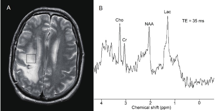
Figure 9.
Proton magnetic resonance spectroscopy used to distinguish radiation necrosis from recurrent tumor. A: Localization of a voxel in a patient with a history of anaplastic astrocytoma treated with surgery and radiation. Most of the area within the voxel demonstrated contrast enhancement on postgadolinium T1-weighted magnetic resonance imaging. B: An elevation of the choline (Cho) peak relative to the creatine (Cr) and N-acetyl aspartate (NAA) peaks and the presence of lactate (Lac) are consistent with recurrent tumor.In an area of radiation necrosis, the peaks of Cho, Cr, and NAA would be markedly reduced. Used with kind permission from the Barrow Neurological Institute.

