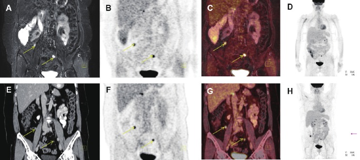
Figure 10.
Coronal PET/MR and PET/CT images showing peritoneal carcinomatosis of a patient with relapse after surgery and chemotherapy for ovarian cancer (yellow arrows). A: T2-weighted STIR MR image, B: FDG-PET from the PET/MR scanner app. 120 min post-injection, C: Fused PET and MR from the PET/MR scanner, D: MIP PET from the PET/MR scanner, E: coronal CT image, F: FDG-PET from the PET/CT scanner (app. 60 min post-injection), G: Fused PET and CT from the PET/CT scanner and H: MIP PET form the PET/CT scanner.

