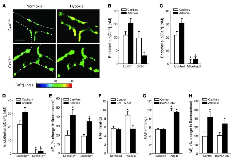
Figure 7. Role of endothelial [Ca2+]i in acute HPV.
(A) Representative images (of 5 replicates) of fura-2–loaded lung arterioles showing endothelial [Ca2+]i at normoxia (21% O2) and after 10 minutes of hypoxia (1% O2) in Cx40+/+ and Cx40–/– lungs. Vessel margins are denoted by dotted lines. Scale bar: 50 μm. Group data (n = 5 lungs each) show endothelial Δ[Ca2+]i in response to acute hypoxia in pulmonary capillaries and arterioles of (B) Cx40+/+ and Cx40–/– mice or (C) Cx40+/+ lungs in the absence (control) or presence of the VDCC blocker mibefradil (10 μM). *P < 0.05 vs. Cx40+/+ or control. Group data (n = 5 lungs each) showing (D) endothelial Δ[Ca2+]i or (E) ΔEm in response to acute hypoxia in capillaries and arterioles of Cacna1g+/+ and Cacna1g–/– lungs. *P < 0.05 vs. Cacna1g+/+; #P < 0.05 vs. capillary. Group data (n = 5 lungs each) showing PAP in isolated perfused Cx40+/+ lungs (F) at normoxia and after 10 minutes of hypoxia or (G) at baseline and after Ang II (1 μg bolus) in control lungs or after endothelial Ca2+ chelation by BAPTA-AM (40 μM). *P < 0.05 vs. normoxia or baseline; #P < 0.05 vs. control. (H) Group data (n = 5 lungs each) showing endothelial ΔEm in response to acute hypoxia in pulmonary capillaries and arterioles in the absence (control) or presence of BAPTA-AM. *P < 0.05 vs. capillary.

