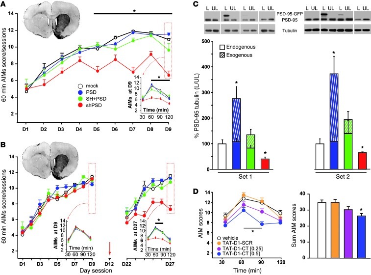
Figure 2. Downregulating PSD-95 in the DA-deprived striatum reduces AIM severity in the hemiparkinsonian rat.
(A) Both the peak AIM scores (rated at 60 minutes) from day 5 as well as the time course AIM (bottom right inset) scores were significantly reduced with shPSD LV compared with mock control. *P < 0.05, shPSD vs. mock. (B) Rats displayed identical peak AIMs after 9 l-DOPA treatment days before receiving LV at day 12 (red arrow). Peak AIM score in the shPSD group was significantly reduced compared with all other groups. This reduction was further highlighted in a time course experiment at day 27 (bottom right insets), with a significant decrease 60 and 90 minutes after l-DOPA administration. In both sets, animals had similar loss of dopaminergic innervation in the striatum as detected by TH immunohistochemistry (top left insets). *P < 0.05, shPSD vs. all other groups. (C) Detection of the endogenous PSD-95 and exogenous PSD-95–GFP transgene by Western blot in striata extracts from 2 sets of animals. Both lesioned (L) and unlesioned (UL) sides are shown. In both experiments, shPSD significantly reduced the expression of endogenous PSD-95, whereas PSD significantly increased PSD-95, compared with the mock group. Lanes were run on the same gel but were noncontiguous (white lines). *P < 0.05 vs. mock. (D) While TAT-D1-SCR peptide had no effect on AIM score, TAT-D1-CT (0.25 and 0.5 nmol) dose-dependently reduced AIM score at peak and at 60 and 90 minutes after l-DOPA administration, resulting in overall improvement. *P < 0.05 vs. vehicle.

