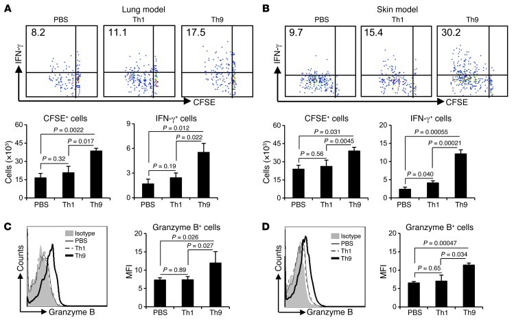
Figure 6. Th9 cell treatment enhances tumor-specific CD8+ CTL differentiation.
(A and C) CFSE-labeled OT-I CD8+ T cells (3 × 10⁶) were i.v. transferred into mice bearing 5-day pulmonary B16-OVA melanoma, which were also i.v. transferred at the same day with 3 × 10⁶ Th1 or Th9 cells. (B and D) CFSE-labeled OT-I CD8+ T cells (3 × 10⁶) were i.v. transferred into mice s.c. injected with 5 × 10⁵ B16-OVA cells, together with s.c. injection of Th1 or Th9 cells at the same site of tumor cell inoculation on the same day. All mice (n = 3/group) were sacrificed 3 days later, and OT-I cells from TDLNs were analyzed by FACS after restimulation. (A and B) Upper panels show the frequency (%) of IFN-γ–producing CFSElo (proliferated) OT-I cells; lower panels show total CFSE+ OT-I cells and total CFSElo IFN-γ–producing OT-I cells recovered from TDLNs. (C and D) Histograms show granzyme B production by CFSE+ OT-I cells; graphs show mean fluorescence intensity of granzyme B expression by CFSE+ OT-I cells recovered from TDLNs. P values are shown as indicated.

