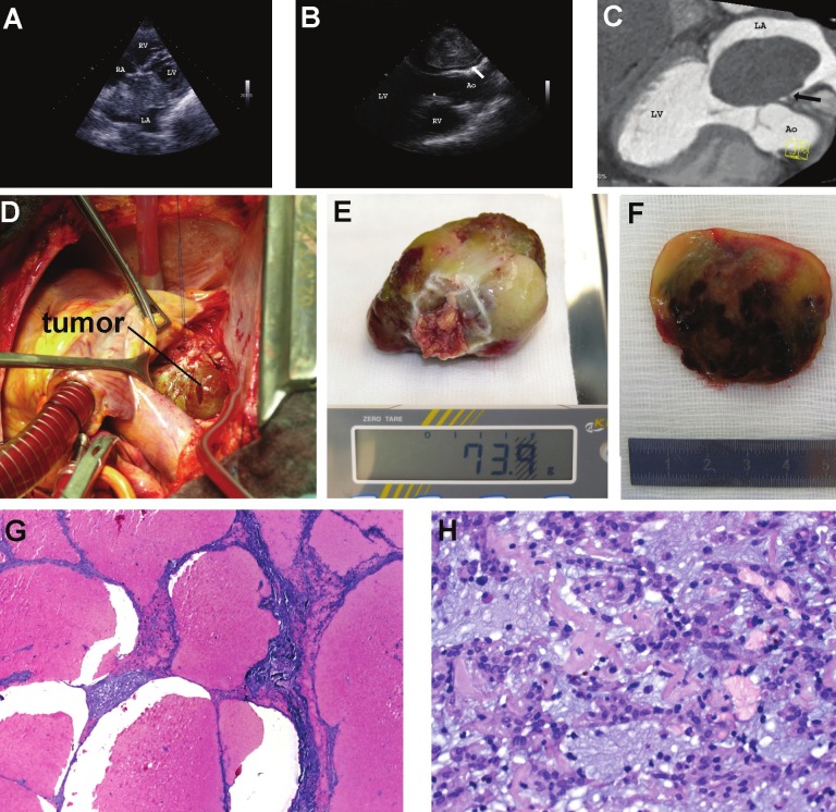
Figure 1.
A: Transthoracic echocardiography (modified 4-chamber view) showed a large mobile left atrial mass (68 × 52 mm) attached to the atrial septum protruding into the left ventricle during diastole (LV, left ventricle; LA, left atrium; RV, right ventricle; RA, right atrium); B: Transesophageal echocardiography (long-axis view) revealed large heterogeneous, lobulated echogenic mass with a short stalk (arrow) attached to the interatrial septum, obstructing diastolic filling of the left ventricle (Ao, Aorta); C: Computed tomographic scan showing large globular mass filling left atrium with narrow base of attachment to the interatrial septum; D: Intraoperative photograph showing the giant left atrial tumor mass; E: The gelatinous tumor formation weight 74 mg; F: The cut section of the resection specimen showed extensive peliosis-like cystic hemorrhagic spaces within otherwise tan-yellow to obviously myxoid tissue; G: Histological examination of Hematoxylin and Eosin (H&E)-stained sections confirmed the presence of large cystic spaces lined by compressed tumorous tissue, but lacking true endothelial lining; H: The tumor showed otherwise typical features of atrial myxoma with ovoid to spindled or rounded bland-looking tumor cells arranged in cords, microtrabeculae, nests and perivascular ribbons within a strikingly myxoid edematous background rich in small-sized capillaries with occasional hemosiderin pigments indicating recurrent stromal bleeding.

