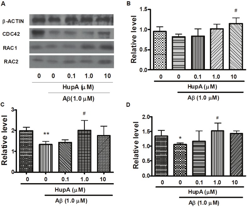
Figure 2.

Western blotting analysis of CDC42, RAC1 and RAC2 protein levels in Aβ treated SH-SY5Y cells. After incubation with PBS, Aβ, HupA with Aβ for 24 h, SH-SY5Y cells were harvested and analyzed by western blot. Figure is the representative bands of β-ACTIN, CDC42, RAC1 and RAC2 Western blotting. A. Representative β-ACTIN, CDC42, RAC1 and RAC2 western blot bands. B, C and D. Immunoblots quantification of CDC42, RAC1 and RAC2, respectively. All bands were quantified and normalized by β-ACTIN. The data is expressed as mean ± SEM from three independent experiments. *p <0.05, **p <0.01 compared to control. #p < 0.05 compared to Aβ-treated group.
