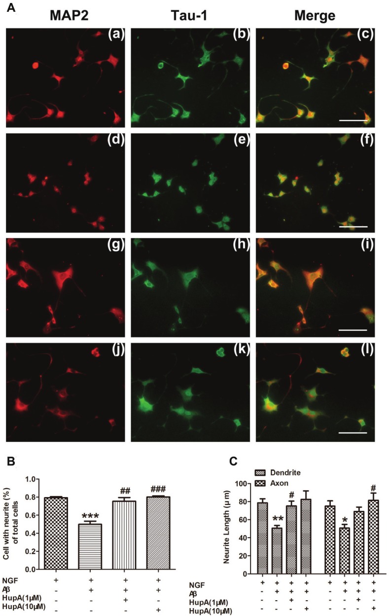
Figure 4.

The effect ofHupA on the NGF-inducedneurite outgrowthof PC12 cells with Aβtreatment. Cells weretreated with NGF (a-c),NGF and 1 μM Aβ1-42(d-f), 1 μM (g-i) or 10μM (j-l) HupA, respectively.HupA was added2h before Aβ treatmentA. shows the representativeimages of immunofluorescentstainingunder different treatments:MAP-2 (red) (a,d, g, j), Tau-1 (green) (b,e, h, k) and merged imagesrevealed both (c, f, i, l). B. Neurite-bearingcells counting. Neuriteformation is measuredunder a fluorescent microscopeafter immunofluorescentstaining.The processes longerthan one cell diameterwere counted as neurites.C. The neuriteslength measurement.The longest length ofneurites was measuredafter immunofluorescentstaining, and themean value of neuritelength was calculated.Values are representedas means ±SEM. *p<0.05, **p <0.01 and ***p <0.001 for comparisonsbetween NGFtreatment and co-treatmentof NGF and Aβ; #p<0.05, ##p <0.01 and ###p <0.001 for comparisonsbetween NGFand Aβ co-treated groupand NGF, Aβ and HuperzineA co-treated group.Scale bar is 100 μm.
