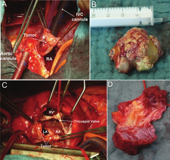
Figure 3.

A: Intraoperative photograph showing large polypoid exophytic angiosarcoma after opening of the right atrium. RA, right atrium; IVC, inferior vena cava. B: Intraoperative gross photograph showing a giant myxosarcoma with yellowish myxoid appearance (maximal diameter of 65 x 40 x 45 mm). C: Intraoperative photograph showing a diffuse tumor recurrence in the left and right atrium attached to the tricuspid valve. D: Resected tumor mass with valve leaflets (same as C). LA, left atrium; RA, right atrium; RV, right ventricle.
