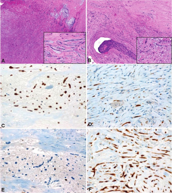
Figure 5.

Cardiac myxosarcoma probably originating within myxoma. A: residual myxoma-like areas (upper right)blended with sclerosing atypical spindle cell sarcoma (lower left). Inset: myxoma-like cells lacked significant atypia.B: focus of venous invasion was seen within adjacent myocardium (Inset: note atypical hyperchromatic nuclei). C:Myxoma-like areas strongly expressed calretinin, as opposed to isolated positive cells in the sarcomatous area (D).On the contrary, p16 was nearly absent in the myxoma-like component (E) but diffusely ands strongly expressed inthe atypical component (F).
