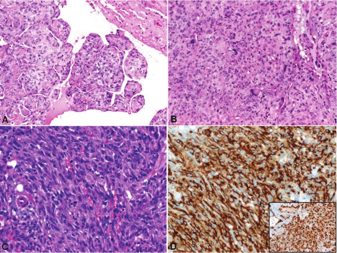
Figure 6.

Example of non-myxoid cardiac sarcomas. A: This undifferentiated sarcoma/MFH showed a papillary polypoid component that was probably responsible for the brain and intestinal metastasis detected later in this patient. B: typical solid growth pattern with pleomorphic multinucleated tumor cells and significant mitotic activity. C: the angiosarcoma displayed deceptive spindle cell morphology. D: strong cytoplasmic expression of CD31 (main image) and nuclear reactivity with ERG (subimage) in the angiosarcoma.
