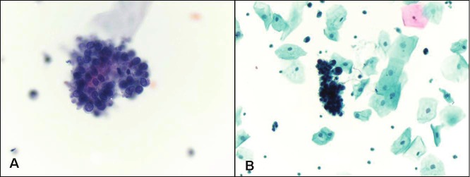
Figure 1.
Low grade urothelial carcinoma. A. Papillary cluster of malignant cells with high nuclear to cytoplasmic ratio (Papanicolaou stain, magnification x 400). B. Three-dimensional cluster of malignant cells with nuclear pleomorphism, hyperchromasia and irregular nuclear borders (Papanicolaou stain, magnification x 200).

