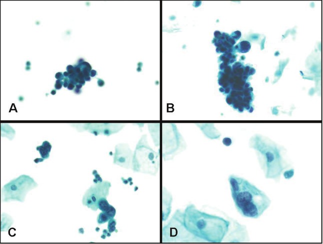
Figure 4.

Case of urothelial carcinoma showing mixed low and high grade features. Note tight clusters of malignant hyperchromatic low grade tumor cell clusters with dense cytoplasm and irregular nuclear borders (in A and B), and rare, isolated clusters of high grade malignant cells (in C and D) (Papanicolaou stain, magnification x 200).
