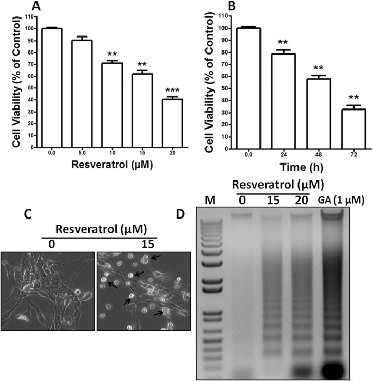
Fig. 1.
Resveratrol increases cellular cytotoxicity of Rat B103 neuroblastoma cells. (A, B) B103 cells were cultured in 96-well culture dishes to near confluence 50~60% and then starved in DMEM containing 0.5% FBS for 24 h. Cell death was determined by using the cytotoxicity assay kit (CCK-8, Dojindo Lab). Cells were exposed to resveratrol in a different dose of 0 to 20 µM in dose dependent and 15 µM in time dependent experiments. Each point is mean±SEM of quintuple samples. Data are mean from three independent experiments in which the activity in the absence of resveratrol versus in the presence of resveratrol is significantly different (n=3, **p<0.01). (C) B103 cells were grown in 24-well culture dishes to near confluence 50% and then starved in DMEM containing 0.5% FBS for 24 h. They were then added 15 µM concentration of resveratrol and grown at 37℃, in humidified 5% CO2 for 48 h and then morphology was observed by Bright-Field Microscopy. Arrows indicate cells with apoptotic morphology. (D) B103 cells were grown in 100 mm culture dishes to near confluence 90% and then starved in DMEM containing 0.5% FBS for 24 h. The cells were then treated with 0, 15 and 20 µM of resveratrol. After 48 h resveratrol treatment, DNA was extracted and separated on 0.8% agarose gel containing ethidium bromide. DNA fragments were visualized under UV light. M indicates as a Marker. GA (Gambogic acid) used as a positive control.

