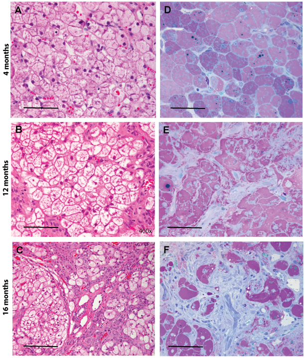
Fig. 3.
Marked glycogen accumulation is present in hepatocytes at 4, 12 and 16 months of age. (A–C) Paraffin-embedded, H&E-stained liver sections illustrate the typical vacuolated appearance of glycogen-filled hepatocytes at 4, 12 and 16 months. (D–F) In HRLM sections stained with PAS-Richardson’s stain, the glycogen is well preserved and appears light purple. Dense fibrosis is evident in F; fibroblasts stain light blue. Scale bars: 50 μm (A,B,F), 100 μm (C) and 30 μm (D,E).

