Fig. 5.
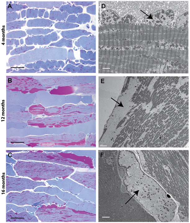
Progressive cytoplasmic glycogen accumulation occurs in skeletal of GSD3 dogs. (A–C) Progressive accumulation of glycogen in skeletal muscle over time. MetaMorph measurements were 6.5±3.1%, 20.3±7.6% and 17.3±4.7% tissue area occupied by glycogen at 4, 12 and 16 months, respectively (HRLM, PAS-Richardson’s stain). (D–F) Ultrastructural changes that occur over time. At 4 months, glycogen accumulates in the cytoplasm and dissects in between myofibrils and just beneath the cytoplasmic membrane, forming small blebs (D). At 12 months, the cytoplasmic glycogen begins to pool and disrupts the contractile apparatus, causing fraying of myofibrils (E). At 16 months, entire regions of cells are filled with glycogen, displacing all contractile elements, leaving only mitochondria to float within the pools of glycogen (F). Arrows indicate glycogen pools. Scale bars: 50 μm (A–C), 2 μm (D), 5 μm (E) and 6 μm (F).

