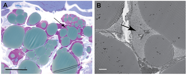
Fig. 6.
Accumulation of cytoplasmic glycogen within adipocytes is apparent in 16 month biopsies. (A) Glycogen stains purple at the periphery of fat globules within adipocytes present in skeletal muscle biopsies (HRLM, PAS-Richardson’s stain). (B) Electron microscopy demonstrates the finely granular ultrastructure of the glycogen surrounding fat globules in adipocytes. Arrows indicate glycogen pools. Scale bars: 30 μm (A) and 5 μm (B).

