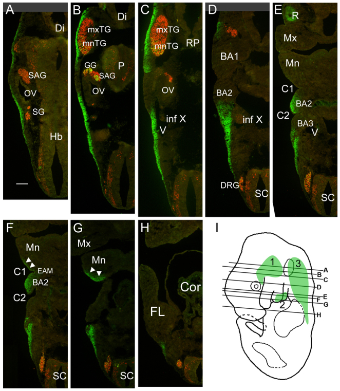
Fig. 2.

Hmx1 expression in developing mouse craniofacial mesenchyme. (A–H) Serial sections of a wild-type E11.5 mouse embryo showing the expression domains of Hmx1 in the CM. Successively more ventral planes of section show distribution of Hmx1-expressing cells in the CM. Arrows in F and G indicate the region of expression in ventral BA1. (I) Planes of section in A–H. Shaded areas indicate regions of Hmx1 expression: 1, part of proximal BA1 overlying the trigeminal ganglion; 2, the caudal half of distal BA2 and a small region of the caudal/distal tip of the mandibular component of BA1; 3, an extensive region of posterior mesenchyme, caudal to the head vein and branchial arches, ending at the top of the limb bud. BA1, branchial arch 1; BA2, branchial arch 2; BA3, branchial arch 3; C1, brachial (pharyngeal) cleft (groove) 1; C2, brachial cleft 2; Cor, heart; Di, diencephalon; DRG, dorsal root ganglion; EAM, external auditory meatus; FL, forelimb; Hb, hindbrain; GG, geniculate ganglion; Inf X, inferior ganglion of the IX–X ganglion complex (nodose/petrosal ganglion); Mn, mandibular process (of BA1); Mx, maxillary process (of BA1); mnTG, mandibular lobe, trigeminal ganglion; mxTG, maxillary lobe, trigeminal ganglion; OV, otic vesicle; P, pons; RP, Rathke’s pouch; SAG, statoacoustic ganglion; SC, spinal cord; SG, superior ganglion; V, head vein. Scale bar: 200 μm.
