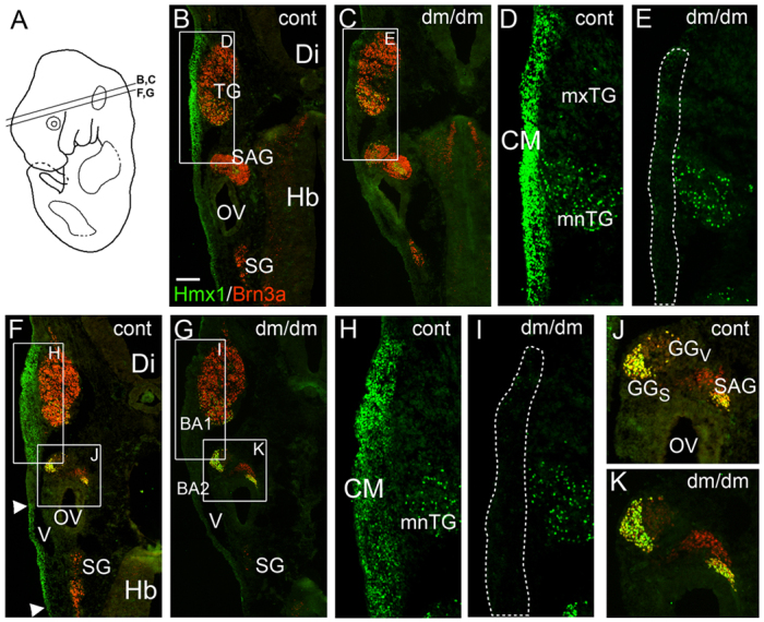
Fig. 3.

Loss of Hmx1 expression in proximal BA1 in the E13 Dumbo rat. E13 rat embryos, developmentally equivalent to E11.5 mice, were examined using immunofluorescence for Hmx1 and for Brn3a, which identifies somatosensory neurons. (A) Plane of section in subsequent views. (B–E) Control (B,D) and dumbo (C,E) embryos showing loss of expression of Hmx1 in the dorsalmost part of BA1 in the mutant. The CM overlying the TG is present (dashed line in E), but fails to express Hmx1. Expression of Hmx1 is unaffected in the mnTG and SAG. (F–K) Control (F,H,J) and dumbo (G,I,K) embryos showing loss of Hmx1 expression in BA1 in dumbo embryos. Some loss of Hmx1 expression is also noted in posterior mesenchyme overlying the head vein in the mutant (arrowheads, F). CM, craniofacial mesenchyme; Di, diencephalon; GG, geniculate ganglion; GGS, geniculate ganglion, somatosensory component; GGV, geniculate ganglion, viscerosensory component; Hb, hindbrain; mnTG, mandibular lobe, trigeminal ganglion; mxTG, maxillary lobe, trigeminal ganglion; OV, otic vesicle; SAG, statoacoustic ganglion; SG, superior ganglion (of IX–X ganglion complex); TG, trigeminal ganglion, V, head vein. Scale bar: 200 μm.
