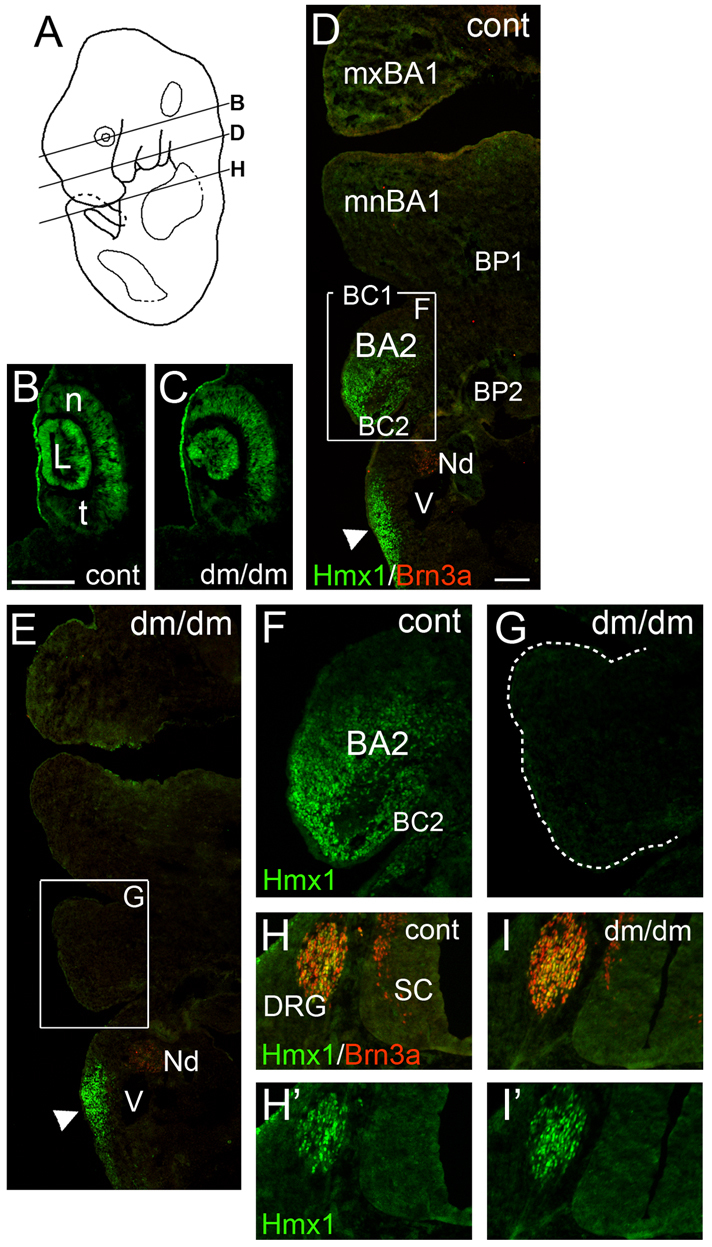
Fig. 4.

Loss of Hmx1 expression in BA2 in the E13 Dumbo rat. (A) Plane of section in subsequent views. (B,C) Hmx1 is expressed in a nasotemporal gradient in the retina which is unchanged in dumbo embryos. (D–G) Expression of Hmx1 in the distal branchial arches; top of figures is anterior. Hmx1 is not expressed in distal BA1 at this stage. Hmx1 expression in distal BA2 is lost in the dumbo embryo (G). Expression of Hmx1 in posterior mesenchyme is not affected (D,E arrowheads). Some neurons in the inferior part of the tenth ganglion are weakly positive for Brn3a. (H–I′) Expression of Hmx1 in the DRG is unchanged in dumbo embryos. Top of figure is dorsal. BA2, branchial arch 2; BC1, brachial cleft 1; BC2, brachial cleft 2; BP1, brachial pouch 1; BP1, brachial pouch 2; DRG, dorsal root ganglion; L, lens; mxBA1, maxillary component of BA1; mnBA1, mandibular component of BA1; n, nasal (aspect of retina); Nd, inferior tenth (nodose) cranial ganglion; t, temporal (aspect of retina); SC, spinal cord; V, head vein. Scale bars: 200 μm (B) and 100 μm (D).
