Fig. 5.

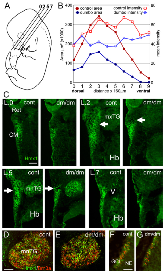
Altered Hmx1 expression in the E14 dumbo rat. Hmx1 expression was examined by immunofluorescence in the lateral cranial mesenchyme of E14 embryos, at which stage the branchial arches are no longer anatomically distinct structures. (A) Planes of section for levels analyzed in subsequent views. (B,C) Semiquantitative analysis of Hmx1 expression in BA1 at E14. The area and intensity of Hmx1 expression was measured at 160-μm intervals encompassing the cranial mesenchyme lateral to the trigeminal ganglion and pons. The overall area of Hmx1 immunofluorescence staining were significantly diminished in the dumbo embryo (paired t-test, n=10, P=0.00017). In the more dorsal sections, the loss of expression occurred mainly rostral to a boundary at about the midpoint of the trigeminal ganglion (arrows). The main effect on Hmx1 expression was to reduce the area of expression, but the intensity of the immunofluorescence signal was also somewhat diminished in the expressing area (paired t-test, n=10, P=0.003). (D,E) Expression of Hmx1 in mandibular lobe of trigeminal ganglion. (F,G) Expression of Hmx1 in differentiating retinal ganglion cells, some of which also express Brn3a. CM, craniofacial mesenchyme; GCL, ganglion cell layer; Hb, hindbrain; mnTG, mandibular lobe, trigeminal ganglion; mxTG, maxillary lobe, trigeminal ganglion; NE, neuroepithelium; Ret, retina; V, head vein. Scale bars: 100 μm (C,D) and 50 μm (F).
