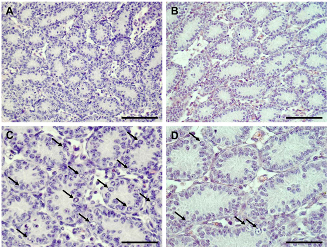
Fig. 1.
Representative histology of WT and SCCx43KO mouse testis at day 8 p.p. H&E staining of a WT mouse testis (A,C) shows SC and type A and B spermatogonia (arrows). By contrast, in seminiferous cords of an SCCx43KO mouse (B,D), only single GCs (arrows) are detectable. Scale bars: (A,B) 100 μm; (C,D) 50 μm.

